
Dental Radiography
The knowledge gained from Dental Radiography not only improves patient care, but more importantly, the information gained via these radiographs speeds dental procedures and decreases complications.
Nearly all veterinary patients have some form of oral disease. Dental radiographs are a critical piece of information for the veterinarian for both diagnosing and treating oral disease.
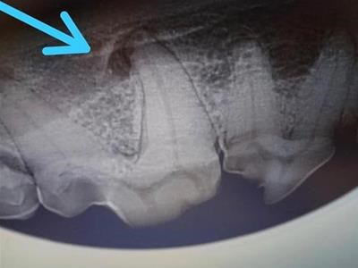
The above image shows what was a healthy tooth to look at upon visual examination, but when xrayed, an abscess was noted on one of the roots.
Cats with holes in their teeth need those teeth to be extracted. Xrays inform us whether they can be crowned (i.e. the top of the tooth cut off) or whether the tooth roots also need to be removed. If the roots are being reabsorbed by the alveolar bone they do not need removal . This saves time to complete the procedure and money for the owner.
We use DR images which are acquired by placing a sensor into the mouth in the same position as a film and exposing the sensor with a greatly reduced dose of radiation compared with traditional dental film. The image is transferred within seconds for viewing on a computer. These images are then electronically stored and manipulated as needed for radiographic evaluation of a wide variety of dental lesions.
Dental radiography is recommended in the evaluation of:
- Tooth resorption
- Periodontal disease
- Endodontic disease, including discolored or fractured teeth and facial swelling
- Retained roots
- Missing teeth
- Abnormally located teeth
- Malformed teeth
- Osteomyelitis
- Boney lysis secondary to neoplasia
- Metabolic bone disease
- Dentigerous cysts (localization)
- Traumatic injuries
Dental radiography is indispensable in the development of an appropriate treatment plan.
Book an Appointment
- If you wish to book your pet in for a dental procedure, please call the practice directly on 01 885 3253 so that we can give you all the necessary information.
- For routine procedures: BOOK ONLINE NOW